Internal structure of the ovary
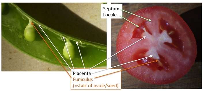
An ovary may have one or more locules (internal spaces or voids in which the ovules reside).
If an ovary contains more than one locule, the walls between locules are called septa.
Ovules are attached to a part of the ovary wall called the placenta. This placenta often looks lighter and less dense that the rest of the ovary wall.
Several patterns of placentation are recognized.
Marginal placentation consists of a vertical line of placenta up one side of a locule in an ovary with a single locule.
Parietal placentation is similar, but consists of two-to-many vertical lines of placenta up the inner walls of an ovary with a single locule. Parietal placentation is found in some ovaries of compound pistils (pistils consisting of more than one fused carpel).
Axile placentation exists in ovaries of compound pistils that have multiple locules. In axile placentation, the placenta exists in the center of the ovary, where all of the septa join.
Free-central placentation consists of a single, central column of placenta in an ovary with one locule. It is thought to have evolved from an ancestor with axile placentation through the evolutionary loss of septa within the ovary.
Basal placentation consists of a lump or very short column of placenta in the bottom of an ovary.
Basal placentation is a very derived pattern of placentation. Whereas marginal placentation, parietal placentation, and axile placentation give good clues as to how many carpels are in a pistil, most of those clues have disappeared in pistils with basal placentation.
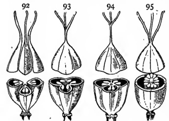
The figure here shows the gynoecium in the flower of four different species. Each gynoecium contains three carpels.
Test your knowledge below.